Terahertz scattering reveals signs of damage and disease in tissues
Stony Brook University method could assist diagnosis of cancer or treatment of burns.
11 June 2025 Research & Development
 A project at New York's Stony Brook University has used terahertz (THz) polarimetry to detect microscopic tissue changes associated with skin damage from burns, a method that may also be suitable for detecting signs of malignancy in skin cancer.
Discussed in Journal of Biomedical Optics, the findings could indicate a new way for clinicians to identify and monitor diagnostic markers, leading to improved therapy and treatment.
The potential value of THz imaging in biomedical applications has been recognized for some time. Falling between the infrared and microwave regions of the spectrum, its relatively non-invasive and non-ionizing nature can give it inherent advantages for imaging of fragile surfaces such as skin tissues.
However, current THz imaging techniques face significant limitations for medical applications, commented the Stony Brook project.
"Most existing approaches rely primarily on water content differences between healthy and diseased tissue as their main source of diagnostic contrast, an approach that proves overly simplistic for complex disease conditions," noted the team.
"Moreover, while polarization measurements of reflected THz waves seem to be valuable for tissue diagnosis, the underlying mechanisms that create different polarization responses in tissues remain poorly understood."
The project's solution was to study how polarized THz light interacts with specific microscopic features that vary between healthy and diseased or damaged tissue. It looked in particular at the behavior termed Mie scattering, occurring when the scattering features have a diameter comparable to the incident wavelength.
Measurements of polarization changes due to Mie scattering from structures within the tissue could be a valuable route to quantifying different degrees of disease progression, noted the team.
THz technology used in routine medical diagnosis
Given the complexity of THz interaction with tissues, Stony Brook first used tissue phantoms to optimize the available modelling of THz waves scattered from spherical particles embedded in highly absorbing biological media. This work showed that the tissue's polarization properties can be characterized using just a single polarization measurement, unlike conventional approaches requiring at least four.
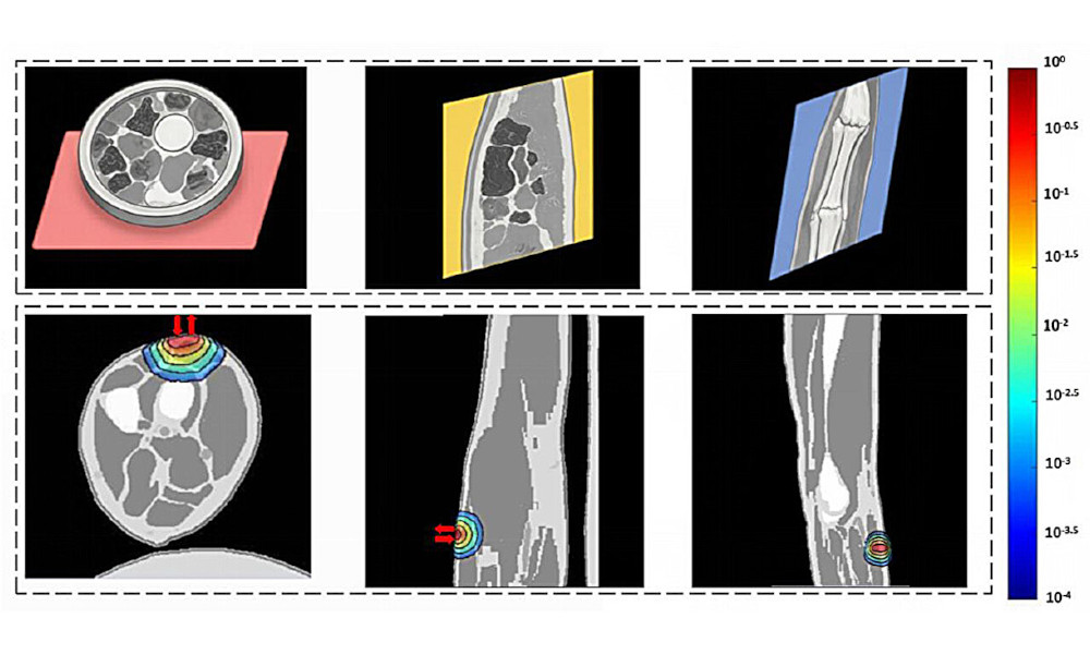
The project then applied its technique to the THz imaging of induced burns in ex vivo porcine skin samples, where mapping the degree of polarization and the diffuse backscattered intensity showed clear contrast between the burned and healthy tissues.
A higher degree of polarization in burned skin may come from the destruction of large skin structures and so an overall decrease in the average size of scattering sources, theorized the team in its JBO paper. The higher backscattered intensity could be the result of water loss from the damaged tissues.
The next steps will involve applying the technique to cancer tissue samples, and expanding the THz measurement capabilities to capture even smaller tissue features. With larger bandwidth, this polarimetric technique could potentially resolve structures as small as 10 to 30 microns in size, encompassing a wider range of disease-related tissue changes.
"As THz technology continues to advance, the results of this study represent a significant step toward its inclusion in routine medical diagnosis, potentially transforming how clinicians detect and monitor disease progression," said the project.


Exail poised for Thales takeover
July 13 2026
Magic Leap pivots to AR waveguide supply
July 13 2026