NSF grant funds AI-powered camera for surgical imaging
University of Tennessee project aims for fully insertable wireless robotic imaging device.
05 November 2025 Research & Development
 A project at the University of Tennessee Medical Center (UT Medical Center) has received a $1 million grant in support of its medical imaging technology.
The funding from the National Science Foundation (NSF) will assist UT's incorporation of AI into its ongoing efforts towards minimally invasive implantable cameras.
The Tennessee lab of Jindong Tan has spent a decade working on novel techniques for implantable cameras that will improve patient outcomes, enhancing the design and function of the imaging device.
"We are trying to find gaps in the marketplace and seeing how we can improve upon either laparoscopic surgery or robotic surgery, so that it's cheaper, safer, or faster than what is currently out there," said Tan.
This work has involved tackling the practical issues arising from an implanted miniaturized imaging system. In 2017, for example, Tan demonstrated how in vivo lens cleaning could be incorporated into a robotic laproscopic camera by creating a carbon dioxide gas barrier in front of the lens.
Other breakthroughs include untethered designs of camera that function without tethering wires, through combinations of wireless video transmission, onboard power sources and Bluetooth control.
The ultimate goal is a camera device free of tethering wires, operated from outside by a robot and inserted through the same incision used for surgery. Avoiding additional incisions helps to minimize the interference and reduced camera mobility that they can bring.
The best surgical outcome possible
"We are talking about less incisions, faster recovery time, less blood loss," Tan said. "Hopefully it's a camera that provides a super vision for surgeons to achieve the best surgical outcome possible. Because during surgery, if you use the regular laparoscope, it could get blurry and folded up and you need to take it out, which delays the surgery."
UT's device has been primarily designed for the abdominal wall or chest area, but Tan's team hopes to expand the scope to areas of the body with tighter spaces such as the nose, small joints, or potentially the brain.
"The imaging technologies are allowing us to be more precise, and if we're more precise then the patient has a better outcome," noted UT Medical Center surgeon Gregory Mancini. "This is going to enhance our ability to do harder, more complicated surgeries with the same, high-quality results."
In designing the imaging device the Tan lab has been driven by the different requirements of surgeons in different scenarios, where a global view or a perspective from certain side angles may be needed. Incorporating AI into the imaging platform will be a key aspect of this flexibility.
The project aims to incorporate images from prior surgeries into the visual information available to surgeons, so that they can use both a live image and a preoperative image together. Once ultimately commercialized, such a functional imaging platform could be further developed by companies for widespread use in surgery.
"The way that AI technology is moving along, this is something we think will have further real-world applications by the end of this project," Mancini said. "That's one of the cool things about it. It's a little less like sci-fi, and more about making it a reality integrated in the imaging system."

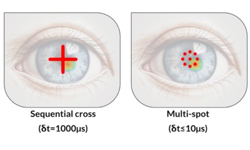
Multi-spot OCT tracks cornea deformations
July 29 2026
Gooch & Housego poised for private equity buyout
July 29 2026