Stanford Medicine maps brain's fiber network at micron-resolution
Computational scattered light method offers new views of neurodegenerative disease.
18 November 2025 Research & Development
 A project at Stanford Medicine has developed a new method of imaging the brain's fiber network, a key structural element in neural function and malfunction.
Every tissue in the human body contains a network of microscopic fibers, shaping how organs function and maintain their structure.
In the brain, degeneration or disruption of this fiber network is related to the decline in neural connectivity found in all neurological disorders. But mapping the fiber architecture with MRI or X-ray techniques has remained challenging.
As discussed in Nature Communications, the new Stanford method is termed computational scattered light imaging (ComSLI), and may point to a simple, low-cost approach for making those hidden structures visible in detail.
"The information about tissue structures has always been there, hidden in plain sight," commented Marios Georgiadis from Stanford's Department of Radiology. "ComSLI simply gives us a way to see that information and map it out."
ComSLI takes advantage of how the orientation of the fiber network influences the scattering behavior of incident light. By rotating the direction of the light and recording how the scattering changes, the direction of the fibers within each microscopic pixel can be reconstructed across the entire image.
The setup requires only a rotating commerical 400-750 nanometer LED light source and a microscope camera, noted the Stanford team, so the approach is accessible and cost-effective compared with other specialized microscopy equipment. Software algorithms analyze the subtle patterns in scattered light to produce color-coded maps, called microstructure-informed fiber orientation distributions, that indicate the orientation and density of fibers within the sample.
Distinct fiber patterns for different tissues
"Spatially oriented structures such as nerve fibers scatter light predominantly perpendicular to their main axis, yielding characteristic light intensity profiles," noted the project in its paper. "ComSLI reverses the light path to determine the orientations of fibers contained in each micrometer-sized image pixel."
In trials ComSLI was applied to samples from the hippocampus, one of the first brain regions affected in many neurodegenerative diseases. Comparing samples from a patient with Alzheimer's disease and a healthy patient showed that the dense fiber crossings normally connecting different parts of the hippocampus were greatly reduced, and one of the main routes for carrying memory-related signals into the hippocampus, the perforant pathway, was barely detectable.
The technique was also used to examine muscle, bone and vascular samples, with each showing distinct fiber patterns related to their physiological roles.
As well as working regardless of how a sample was prepared, ComSLI can also be used to re-analyze slides originally made for other purposes, including those that have been stored for decades. Researchers could then extract structural information from tissue without additional processing.
The ability to map fiber orientation in various species, organs and decades-old specimens could reshape how scientists approach questions of structure and function, said Stanford Medicine. It also turns millions of archived slides worldwide into potential sources of new data.
"This is a tool that any lab can use," said Stanford's Michael Zeineh. "You don't need specialized preparation or expensive equipment. What excites me most is that this approach opens the door for anyone, from small research labs to pathology labs, to uncover new insights from slides they already have."

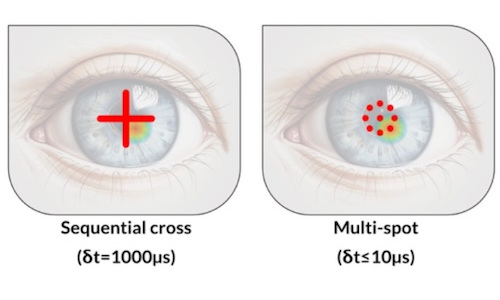
Multi-spot OCT tracks cornea deformations
July 29 2026


Gooch & Housego poised for private equity buyout
July 29 2026