Fluorescence lifetime technique offers real-time imaging of tumor margins
University of California, Davis combines imaging and machine learning to accelerate diagnosis.
17 February 2020 Research & Development
 Accurate imaging of the margins of tumors is a long-standing challenge in breast cancer surgery, where a significant number of patients eventually require a second procedure to remove malignant cells remaining after an initial operation.
Optical techniques are a potentially powerful way to tackle this problem, through Raman spectroscopy, OCT or photoacoustic techniques. But each method has some potential drawbacks, and none has yet achieved wide clinical adoption.
A project at the University of California, Davis has now demonstrated that a combination of fluorescence lifetime imaging (FLIM) and machine learning could potentially allow diagnostic data to be gathered from breast tumor specimens in real-time. The work was published in Biomedical Optics Express.
The breakthrough comes from the UC Davis lab of Laura Marcu, where FLIM techniques and their application to biomedical imaging have been a topic of research for some time.
"Autofluorescence lifetime measurements can overcome the limitations encountered by conventional intensity measurements, such as the variation in fluorescence excitation-collection geometry and attenuation of light absorption by blood within tissue," wrote Professor Marcu in 2018.
"However, to measure and analyze the autofluorescence signal, lifetime-based techniques require complex instrumentation and computational methods that have hampered clinical adoption. There is a compelling need to accelerate the adoption of autofluorescence lifetime techniques that have the potential to improve the surgical outcome and the patient's quality of life."
The new project was designed to develop two particular capabilities vital to this accelerated adoption of FLIM: real-time tissue diagnosis based on parameters derived from fluorescence decay, and an intuitive visualization of tissue type to distinguish between tumor, adipose, and fibrous tissue.
Real-time clinical use on patients
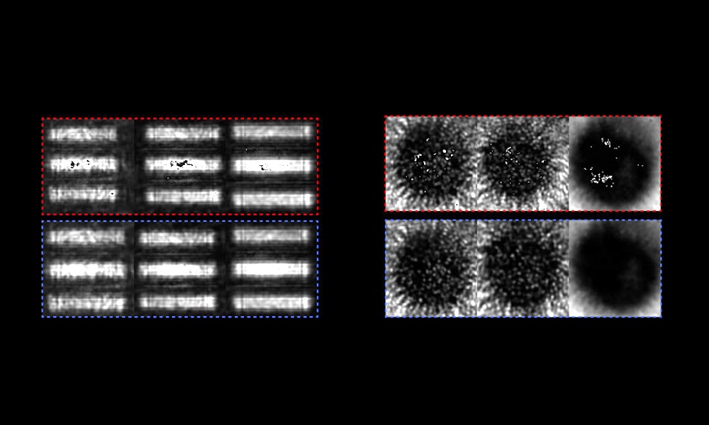
A prototype time-domain multi-spectral time-resolved fluorescence spectroscopy (ms-TRFS) platform developed by the team was used in the study, employing 355-nanometer excitation and spectrally resolving the autofluorescence produced into four spectral bands, so as to detect collagen, porphyrins, and specific dineuleotides of interest.
A numerical method termed a random forest classifier, in which a large number of individual decision trees operate as an ensemble during a machine learning operation, was then applied to the data from a group of 18 patients. The project found that its technique could delineate the cancerous regions in breast specimens with 89 percent sensitivity and 93 percent specificity.
Although the work focused on excised tissue specimens, the ultimate goal is to establish FLIM as an intraoperative tool, able to scan a patient with a fiber optic. According to the team, this will require some adaptations of the procedure, including the creation of volumetric information from a stereo camera set-up, along with a more robust tracking procedure to control the aim of the laser.
But the UC Davis study already demonstrates the potential application of FLIM for margin assessment of breast lumpectomy specimens when combined with machine learning and real-time visualization, according to the group.
"This technique provides high diagnostic prediction accuracy, rapid acquisition, adaptive resolution, nondestructive probing, and facile interpretation of images, thus holding potential for clinical breast imaging based on label-free FLIM," concluded the project team.
Trumpf hopeful of a recovery as orders tick up
July 28 2026
Computational optics startup Elio raises $21M
July 28 2026