University of Nottingham endoscope sees gastrointestinal cancers
Minaturized probe uses spatial frequency domain imaging to spot malignancy.
07 February 2024 Research & Development
 A project at the University of Nottingham has developed an endoscopic imaging platform intended to identify early stage gastrointestinal cancers.
As published in Journal of Biomedical Optics the device is based around spatial frequency domain imaging (SFDI), a promising technique for differentiating between healthy and malignant tissue.
SFDI offers a way to improve the low contrast image quality that endoscopic systems can be prone to, by directing sinusoidal patterns of light at a tissue surface at multiple spatial frequencies and measuring a frequency-dependent tissue response.
Previous applications of this principle to cancer diagnosis have included a project at the University of Buffalo that used the optical absorption and scattering data generated to assess light propagation and attenuation in the living tissue, and see how these were affected by the presence of cancer.
Moves to commercialize devices based on the SFDI principle have also been made, such as the Clarifi imaging system developed by Modulim, formerly Modulated Imaging, and targeted at detecting the signs of diabetes, vascular and kidney diseases.
The Nottingham project set out to develop a SFDI platform able to be deployed endoscopically for the detection of early stage gastrointestinal cancers, with a primary challenge being the need to miniaturize the optics involved.
"Existing systems are not fit for routine endoscopic deployment in the gastrointestinal tract," commented Jane Crowley from the University of Nottingham. "They either use digital micromirror device-based projectors, which are costly and cannot be sufficiently miniaturized; or use fiber bundles which produce low-quality patterns at a limited set of wavelengths and only record low-resolution images; or use rigid endoscopes that are not flexible enough."
Imaging during minimally invasive endoscopic procedures
The project's solution is an ultra-miniature SFDI system using a custom-made bundle of seven optical fibers as a projector, with each fiber independently coupled to laser sources delivering 515 and 660 nanometer light.
"By feeding a single laser of a given wavelength to two different fibers, one can leverage the phenomenon of interference to project a 2D sinusoidal pattern onto the target tissue," said the project. "The spatial characteristics of the resulting pattern can be tuned by selecting different fiber pairs, and patterns consisting of up to three different wavelengths could be projected simultaneously."
This fiber optic system was combined with an ultra-miniature camera measuring 1 millimeter x 1 millimeter and recording 320 x 320 pixel images, to make a prototype SFDI system 3 millimeters in diameter. A custom algorithm tracked phase deviations in the projected sinusoidal patterns, so as to reduce noise in the absorption and scattering profiles being measured.
In trials on phantoms mimicking healthy and cancerous tissues, the prototype provided excellent image contrast between the two types, according to the project. Its specificity and sensitivity for detecting squamous cell carcinoma were estimated to be over 90 percent, said to be on par with the current clinical standards of medical devices.
The next steps could see the device miniaturized further, perhaps to below 1.5 millimeters in diameter, suitable for minimally invasive endoscopic procedures. The design's compatibility with multi-wavelength imaging mean it might be able to capture optical information at different tissue depths, or analyze multiple tissue layers at the same time.
"Our prototype shows promise as a cost-effective, quantitative imaging tool to detect variations in optical absorption and scattering as indicators of cancer," said Jane Crowley. "This work could form the basis of new devices suitable for cost-effective endoscopic deployment for screening of gastrointestinal cancers."
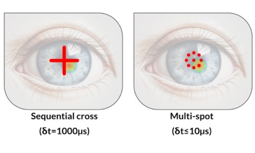
Multi-spot OCT tracks cornea deformations
July 29 2026
Gooch & Housego poised for private equity buyout
July 29 2026

Trumpf hopeful of a recovery as orders tick up
July 28 2026