Wearable microscope reveals details of mouse spinal cord
Salk Institute device could "revolutionize the study of pain."
28 March 2023 Photonics Applications News
 Fluorescence imaging can reveal details of neuronal activity in sections of the spinal cords of animals, but collecting data from living subjects can be challenging.
Developments in micro-optics components such as gradient-index lenses have enabled optical platforms to be greatly miniaturized, but such components can impose their own limitations on the data that can be collected.
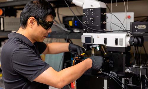
A project at the Salk Institue for Biological Studies has now demonstrated a wearable microscope with customized micro-optics intended to deliver high-speed multi-color imaging, able to be implanted into mice and observe neuronal activity in the spinal cord.
Reported in Nature Communications and Nature Biotechnology, the instrument is said by the project to addresses multiple challenges of previous wearable microscopes, including their limited working distance, resolution, contrast, and achromatic range.
"These new wearable microscopes allow us to see nerve activity related to sensations and movement in regions and at speeds inaccessible by other high-resolution technology," said Axel Nimmerjahn of the Salk Institute's Waitt Advanced Biophotonics Center. "Our wearable microscopes fundamentally change what is possible when studying the central nervous system."
The device's design goals included a working distance greater than 2 millimeters, spatial resolution of 1 to 2 microns, and a broad achromatic range. After optical modelling, the Institute constructed a pair of instruments built around six custom microlenses, housed in barrel assemblies approximately 7 and 14 millimeters wide and weighing less than 0.2 grams.
A microprism implant was also placed near the tissue regions of interest, a key component of the system according to the project. The microprism increases the depth of imaging so previously unreachable cells can be viewed for the first time, and allows cells at various depths to be imaged simultaneously and with minimal tissue disturbance.
In trials on living mice, the combination of implanted microprism and wearable microscope was able to image astrocytes, glial cells in the spinal cord involved in pain processing, and follow their involvement in sending coordinated signals across spinal cord segments when the tails of the mice were squeezed. Until now it was impossible to know what such cellular activity looked like across those spinal cord regions of moving animals.
"Being able to visualize when and where pain signals occur and what cells participate in this process allows us to test and design therapeutic interventions," said the Salk Institute's Daniela Duarte. "These new microscopes could revolutionize the study of pain."
Duke University computational microscope merges multiple viewpoints
 An image-stitching technology developed by Duke University could also represent a step forward for collecting 3D imaging data from freely-moving living animals.
Described in Nature Photonics, the Multi-Camera Array Microscope (MCAM) represents a new approach to the acquisition of sequential image snapshots to observe large areas or measure 3D information, according to the team.
The technology is being commercialized by a Duke spin-out, Ramona Optics.
MCAM employs a synchronized array of 54 lenses to capture high-speed 3D topographic videos over a 135-sqaure-centimeter area, achieving an imaging speed of up to 230 frames per second. The resulting data, potentially more than 5 gigapixels per second, is then processed by a 3D reconstruction algorithm named 3D-RAPID.
"We've developed new algorithms that can efficiently handle these extremely large video datasets," said Kevin Zhou, lead author of the paper.
"Our algorithms marry physics with machine learning to fuse the video streams from all the cameras and recover 3D behavioral information across space and time."
The platform was used to image zebrafish exposed to neuroactive drugs, looking for changes in behavior that could help researchers develop new potential treatments or better understand existing ones. MCAM allowed researchers obtain the data from live fish, rather than animals that were restrained or knocked out.
"With the 3D and fluorescent imaging capabilities of this microscope, it could change the course of how a lot of developmental biologists do their experiments," said Duke University's Jennifer Bagwell.

Gooch & Housego poised for private equity buyout
July 29 2026

Trumpf hopeful of a recovery as orders tick up
July 28 2026
Computational optics startup Elio raises $21M
July 28 2026