Duke University captures images of curved samples in single snapshot
PANORAMA microscope holds submicron focus across curved or uneven surfaces that span centimeters.
24 September 2025 Research & Development
 A project at Duke University has developed a new microscope platform that can acquire extremely large, high-resolution pictures of non-flat objects in a single snapshot.
Described in Optics Letters, the innovation could speed up medical research and diagnostics, or be useful in quality inspection applications.
The team's goal was to address the inherent challenges involved in imaging samples which are uneven or curved rather than flat, normally limited by issues of field of view, resolution, and imaging speed.
These often compel the use of mechanical scanning to move a biological sample so as to keep various parts in focus, further slowing down the imaging process and creating a bottleneck in modern clinical imaging workflows.
"Although traditional microscopes assume the sample is perfectly flat, real-life samples such as tissue sections, plant samples or flexible materials may be curved, tilted or uneven," said Roarke Horstmeyer from the Duke University Computational Optics Lab.
"With our approach, it's possible to adjust the focus across the sample, so that everything remains in focus even if the sample surface isn't flat, while avoiding slow scanning or expensive special lenses."
The team's new platform is named PANORAMA, or Parallel Adaptive Non-scanning Optical Re-imaging using a Multi-camera Array, and is built around a flat array of 48 micro-cameras, with each focused onto a portion of a curved image plane and relaying the optical signal to a flat sensor array.
This builds on earlier work at the Computational Optics Lab into 3D imaging with an array of microcameras, as a way to meet the need for better 3D imaging available to clinicians during live surgery.
Because each camera can be independently focused to match the sample surface, PANORAMA's entire field of view stays sharp even if the sample is curved and eliminates the need for scanning - essentially a form of tailored adaptive optics. The images from each camera are computationally stitched together into a continuous picture, a process that takes about 5 to 10 minutes according to Duke University.
Huge jump in throughput and flexibility
"The telecentric lens makes it possible to image a very wide field without distortion, while the multi-camera approach overcomes the usual size-and-resolution limit of a single sensor," commented Duke University's Haitao Chen. "This combination lets us acquire a seamless, gigapixel image in a single snapshot, flattening out any curvature adaptively."
In trials using a prepared slide of rat brain tissue under brightfield illumination, the 48-camera array captured the entire slice in one snapshot without any scanning, a 630 MP image. This clearly showed cellular structures measuring as small as 0.84 microns, as well as neurons and dendrites across the sample.
Duke University also used the microscope to simultaneously acquire a brightfield and fluorescence image of onion skin laid over a gently curved surface. When it focused each camera on the local curvature, the entire curved layer stayed sharp. The brightfield images revealed crisp cell walls, while the fluorescence images clearly showed stained nuclei.
The next steps could include adding more cameras or larger sensors to capture an even bigger field in a single shot, perhaps an entire petri dish, along with an automated focus system so each camera no longer has to be adjusted manually for every sample. Computational advances could also enable 3D reconstructions, real-time depth maps or live video of microscopic processes.
"In practical terms, we saw a huge jump in throughput and flexibility: no more moving parts, no tedious focus-stacking, and no blind spots between cameras," said Roarke Horstmeyer. "Compared to older multi-camera microscopes that needed scanning to fill gaps and maintain focus, our approach gives continuous full coverage at sub-micron resolution."

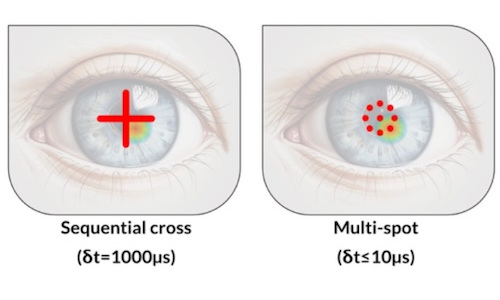
Multi-spot OCT tracks cornea deformations
July 29 2026
Gooch & Housego poised for private equity buyout
July 29 2026