Cryo-optical microscopy freeze-frames cellular activity
University of Osaka technique visualizes cell dynamics during transient processes.
27 August 2025 Research & Development
 Fluorescence microscopy reveals cellular morphology and dynamics in remarkable detail, but achieving clear visualization at fast acquisition rates remains a challenge.
A fundamental trade-off between temporal resolution and how much light can be collected for an image restricts the spatial detail with which fast dynamic cellular events can be captured, meaning important features are lost in dim noisy images.
A project at the University of Osakaa has now developed a cryo-optical microscopy technique designed to take a high-resolution, quantitatively accurate snapshot at a precisely selected timepoint in dynamic cellular activity.
Published in Light: Science & Applications, the findings could provide detailed insights into sample dynamics with improved spatial resolution and temporal accuracy in observations.
"Instead of chasing speed in imaging, we decided to freeze the entire scene," commented Kosuke Tsuji from the University of Osaka.
"We developed a special sample-freezing chamber to combine the advantages of live-cell and cryo-fixation microscopy. By rapidly freezing live cells under the optical microscope, we could observe a frozen snapshot of the cellular dynamics at high resolutions."
Although the potential benefits of cryofixation over chemical fixation in a fluorescence microscopy operation are understood, the optical properties of some fluorescent probes can differ under cryogenic conditions compared to room temperature or may show temperature dependencies, complicating the imaging operation.
The project's optical platform employed four microscopy modalities: a widefield fluorescence microscope; a structured illumination microscopy (SIM) system; a hyper-spectral slit-scanning fluorescence microscope; and a multimodal system combining SIM with slit-scanning Raman microscopy.
Intricate cellular information from multiple perspectives
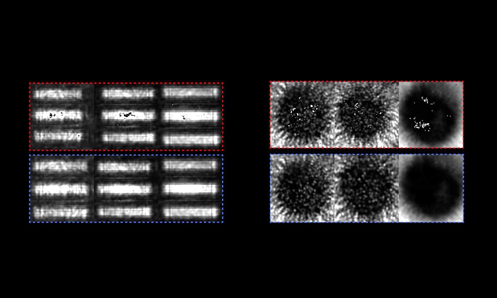
By combining UV light stimulation to induce calcium ion waves with an electrically triggered cryogen injection system, the system could freeze the calcium ion waves at a specific time point after the initiation of an event with 10 ms precision. This let the team arrest transient biological processes with unprecedented temporal accuracy.
The project also studied how to combine its different imaging techniques, which can be difficult to align in time. A near-instantaneous freezing of samples means that multiple imaging modalities can be applied sequentially without worrying about temporal mismatch.
In its study, the project combined spontaneous Raman microscopy and super-resolution fluorescence microscopy on the same cryofixed cells, giving it a view of intricate cellular information from a number of perspectives at the exact same point in time.
Trials on samples including HeLa cancer cells confirmed that the morphological, calcium ion and chemical dynamics of the cells were fixed and imaged successfully by this cryo-optical method. Data related to otherwise intangible information such as pH and redox state could also be detected, using functional fluorescent probes and retrieved from the cryo-fixed cells.
"This research began with a bold shift in perspective: to arrest dynamic cellular processes during optical imaging rather than struggle to track them in motion," said Osaka's Katsumasa Fujita. "We believe this will serve as a powerful foundational technique, offering new insights across life-science and medical research."

Trumpf hopeful of a recovery as orders tick up
July 28 2026
Computational optics startup Elio raises $21M
July 28 2026