Imaging ocular surface assists treatment of dry eye disease
AdOM platform directly images the muco-aqueous tear layer with nanometer resolution
07 October 2019 Photonics Applications News
 A new non-invasive optical imaging system could help to improve diagnosis and treatment of dry eye disease, a condition arising when the inner layer of the tear film protecting the outside of the eye becomes unstable and causes irritation or blurred vision.
Diagnosis of the condition currently relies heavily on patients answering questions about their discomfort, but an imaging platform developed by Israel's AdOM Advanced Optical Methods could lead to more objective visual information about the tear film and its stability. The work was published in Applied Optics.
"Up to 60 percent of ophthalmology office visits are due to dry eye, pointing to the need for a non-invasive and highly accurate device for diagnosis in the office setting," said Yoel Arieli from AdOM. "Our Tear Film Imager (TFI) is the first device that can be used in the ophthalmology or optometry setting to image the tear film and distinguish its inner layers with nanometer resolution."
The new instrument uses an eye-safe halogen light to illuminate the eye, and then analyzes the full spectrum of light reflection. These spectral measurements are used to reconstruct the structures found in the front of the eye, allowing accurate measurement of the tear film inner layers, especially the aqueous sublayer that plays an important role in dry eye but has been difficult to analyze with other methods.
"The broadband illumination source and fine details available from spectral analysis provide nanometer-level insight into subtle changes in each tear film layer and sublayer," said Arieli. "These measurements are completed automatically in just 40 seconds."
Previous strategies for imaging the tear film have employed a number of approaches, including methods based on existing types of keratoscopy, or measurement of the curvature of the cornea. Imaging the reflection of concentric rings projected onto the cornea has been to used as a means of measuring the dimensions of the cornea, but if carried out dynamically as videokeratoscopy, it can also provide information about the behavior of the tear film.
In 2018 Quantel Medical released its LacryDiag platform, in which light from white, blue and infrared LED sources was used to analyze different constituent layers of the tear film.
Clinical trials
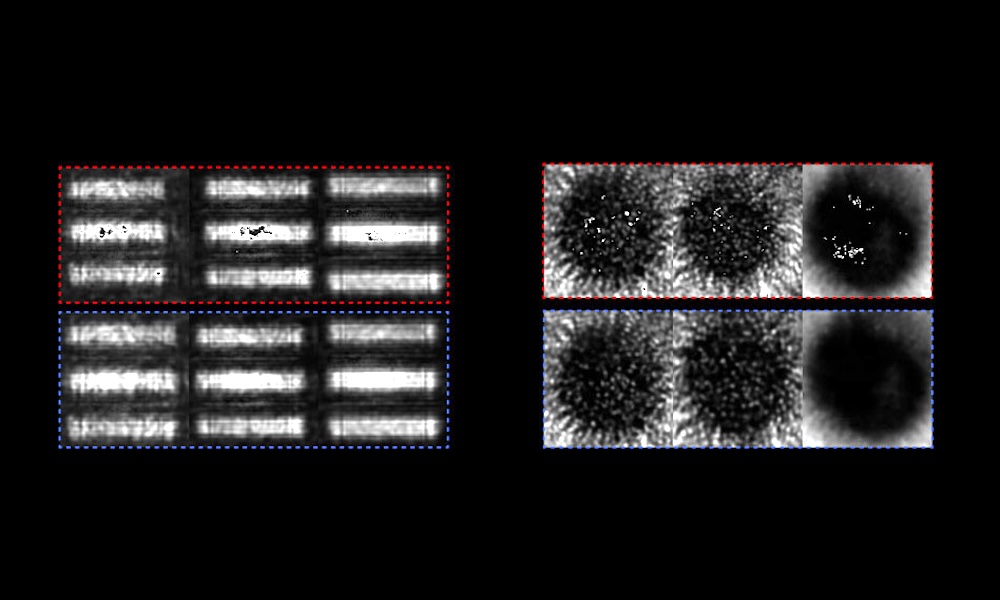
According to AdOM, its new device operates by acquisition of large field-of-view imagery and fast spectrometric measurement of the interference from the thin tear film sublayers. This allows it to generate quantified information about muco-aqueous layer thickness, lipid layer thickness, thickness change rate, and the time taken for the tear film to break up.
"Analyzing the tear film structure is an extremely tough challenge," commented AdOM in its technical description of the new system. "Each inner layer is extremely thin and the subject's eyeball is constantly moving. The TFI tomography capabilities enable 3D imaging as a function of time between blinks to evaluate the behavior of each of the tear film inner layers. Such capabilities can deduce the evaporation rate and uniformity of each layer which is then able to support the physician's decision as to the appropriate care."
After demonstrating a resolution of 2.2 nanometers on a mock tear film, the researchers tested the instrument's ability to take measurements on the human eye with no intervention while the patient was blinking.
The TFI was then used in two clinical studies, in Israel and Canada, examining dry eye diagnosis with the device and dry eye treatments, which can be precisely evaluated with the imager. Larger studies with more diverse groups of patients to set base levels for both healthy eyes and people with dry eye are now planned, as a path towards eventual regulatory approvals.
"The device worked impressively and presents no risk because it is non-invasive and uses a simple light source," said Arieli. "It not only measured the tear film consistently including blinks every few seconds, but the measurements correlated well with other partially invasive, established dry eye diagnostic techniques."

Trumpf hopeful of a recovery as orders tick up
July 28 2026
Computational optics startup Elio raises $21M
July 28 2026