Polarization-based analysis points to quicker detection of prostate cancer
Technique from Aston University and partners exploits anisotropy of proteins to spot signs of illness.
04 September 2024 Research & Development
 An international project led by the UK's Aston University has developed a novel method of detecting biomarkers from cancer in blood samples, an approach termed liquid biopsies.
Published in Nature Scientific Reports, the technique could prove crucial for identifying significant differences between healthy and cancerous samples, according to the project.
The method, developed at the University's Aston Institute of Photonic Technologies under Igor Meglinski, uses a new polarization-based image reconstruction technique to analyze polycrystalline structures in dried blood samples.
These proteins in blood change their shape and how they fit together during the early stages of diseases like cancer.
This characteristic allowed the team to use changes in both the proteins' unique 3D tertiary structure and in the way they join together into quaternary structures to detect and classify the different cells.
"Our study introduces a pioneering technique to the liquid biopsy domain, aligning with the ongoing quest for non-invasive, reliable and efficient diagnostic methods," commented Igor Meglinski.
The breakthrough also joins recent parallel advances in optical detection of cancer biomarkers. In 2023 spectroscopy developer Dxcover described the use of AI algorithms to analyze the infrared spectra of patient blood samples and detect the presence or absence of cancer-indicating species, a method the company intended to apply to eight different types of cancer.
Immense potential for revolutionizing cancer diagnosis
Aston's technique makes use of the Mueller matrix, an approach to mathematical modelling which characterizes the optical properties of a sample by the interaction of polarized light in the absence of non-linear effects.
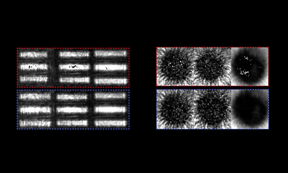
Data from digital cameras can already be used to capture 2D Mueller matrix images, ie. 2D distributions of the elements of Mueller matrices, which can reveal information about the polarization properties at each point. The new breakthrough involved the development of novel 3D Mueller matrix (3D MM) polarimetry techniques using an 633-nanometer interferometer, which can provide 3D information about the proteins being imaged.
The project applied its 3D MM modelling to blood film samples collected from both healthy and diseased volunteers, and developed an analytical protocol for distinguishing between the 3D MM data from healthy tissues and those from abnormal cancerous prostate tissues.
This involved quantitative evaluation of optical anisotropy maps from the sample, characterizing parameters such as birefringance and dichroism distributions and identifying the significant differences in these parameters between healthy and cancerous samples.
Results demonstrated "an exceptional accuracy rate of over 90 percent for the early diagnosis and staging of cancer," according to the project, surpassing existing screening methods.
"This breakthrough opens new avenues for cancer diagnosis and monitoring, representing a substantial leap forward in personalized medicine and oncology," commented Igor Meglinski. "It holds immense potential for revolutionizing cancer diagnosis, early detection, patient stratification and monitoring, thereby greatly enhancing patient care and treatment outcomes."

Trumpf hopeful of a recovery as orders tick up
July 28 2026

Computational optics startup Elio raises $21M
July 28 2026