



University of Arizona redesigns endoscope for detection of ovarian cancer
Improved fluorescence imaging and cell collection offer route to effective screening.
07 April 2026
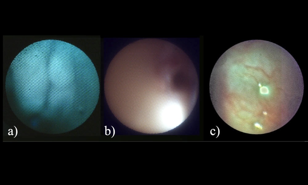
Fallopian tube images using autofluorescence, color white light reflectance and white light reflectance. Credit: Biophotonics Discovery.
A project at the University of Arizona has designed an endoscope intended to assist early detection of ovarian cancer.
Described in Biophotonics Discovery, the project's goal was a submillimeter endoscope combining high‑resolution imaging with gentle cell collection.
Ovarian cancer remains the deadliest gynecologic cancer, noted the Arizona team, largely because it is rarely found early.
Recent research has led to better understanding of how many aggressive ovarian cancers begin, pointing not to the ovary itself but to the fallopian tubes (FT). This in turn has created a need for tools that can safely examine the narrow fallopian tubes structures for early changes linked to cancer.
In 2021 the University of Arizona demonstrated a flexible and steerable submillimeter-diameter endoscope capable of multispectral reflectance imaging, multispectral fluorescence imaging and guided cell collection in the fallopian tubes.
That cell-acquiring fallopian endoscope (CAFE) was based around a 3000-element image fiber bundle with an attached gradient index (GRIN) lens, an architecture which the project noted was successful but with room for improvement in multiple areas of performance.
The CAFE device has now been redesigned and improved in Arizona's Tissue Optics Lab, to increase flexibility and resolution, decrease working distance and weight, and allow color imaging.
The improvements involved combining white‑light LED imaging for navigation with blue‑light reflectance and fluorescence imaging to probe tissue properties, via 405 and 642-nanometer light sources.
A higher‑density fiber bundle and a custom close‑focus lens allowed the system to form sharp images at very short distances where the tube wall lies close to the endoscope tip, noted the team.
Testing in human fallopian tubes
The endoscope also includes a working channel for cell collection. Instead of scraping tissue with an exposed wire, the system uses a smooth, scoop-style collection method that gathers epithelial cells directly into the endoscope tip, reducing the risk of tissue injury.
In trials at NewYork-Presbyterian Queens hospital the redesigned endoscope successfully entered the fallopian tubes of patients, and produced images at multiple locations along their length.
The multispectral imaging capability allowed white-light images to reveal structural features and small blood vessels, while blue‑light imaging captured both reflectance and natural fluorescence signals. Data showed that strong fluorescence signal levels were recorded even with short exposure times, indicating that the system is sensitive enough to detect subtle optical differences in the fallopian tissues.
Researchers analyzed ratios between reflectance and fluorescence signals, as well as color channel ratios in the white‑light images. Measurements were generally consistent between the left and right tubes of the same patient, suggesting that the signals reflect real tissue properties rather than imaging artifacts.
"We were able to show that the endoscope can enter and image inside the fallopian tubes, collect reflectance and fluorescence data, and collect epithelial cells," wrote the project in its paper.
"Continued studies will aim to characterize the variation in fluorescence and reflectance signals from benign FT, as well as examine FT that harbor cancer and other pathologies, to determine if imaging can be used to distinguish benign from pathologic tissue."
University of Nottingham endoscope sees gastrointestinal cancers
February 07 2024

Invisible tattoos that appear when illuminated
October 15 2026

Xiver picks up €28M debt finance to aid photonic chip production
October 15 2026

U.S. Army Research Office awards $19.2 million to Florida team
October 14 2026
ASML looks to add more capacity as chip boom continues
October 14 2026