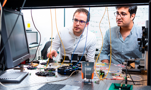



UC Berkeley multimodal platform shows details of subcellular dynamics
Device integrates multiple techniques with adaptive optics.
27 May 2026
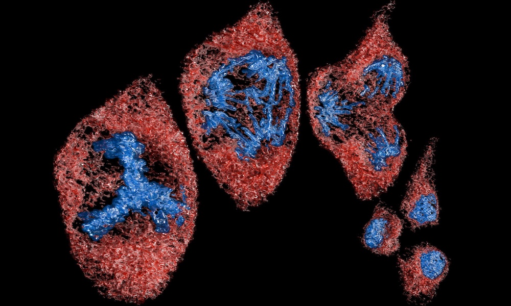
A sequence of high-resolution images showing a cell dividing into three daughter cells, a rare event captured by the MOSAIC microscope in 5D. The images come from the first 3D videos of such an event, which was captured in cancerous pig epithelial cells. Credit: Advanced Bioimaging Center/UC Berkeley.
UC Berkeley's Advanced Bioimaging Center (ABC) has developed a microscopy platform designed to offer new views of cells and embryos, and is using it to train an AI model for living biological systems.
MOSAIC, or Multimodal Optical Scope with Adaptive Imaging Correction, integrates multiple advanced imaging techniques including light-sheet, label-free, super-resolution and multiphoton microscopy, all equipped with adaptive optics.
The modern diversity of advanced microscopy modalities has been driven by the variety of living process and systems being imaged, commented the project in its Nature Methods paper. But optimization for one modality or one class of samples invariably comes with constraints that compromise the study of other systems.
MOSAIC was developed in response, as a single microscope that reconfigures on demand to different imaging modalities, each optimized for a different class of specimens.
The device was first conceptualized as a development of lattice light-sheet microscopy, developed by UC Berkeley's Eric Betzig, with adaptive optics incorporated to improve image quality. MOSAIC has now evolved into a platform with three microscope objectives: for excitation, detection and epifluorescence viewing. The two 1.0-NA detection objectives enable high-resolution, millimeter-scale field-of-view investigations across a variety of imaging modes, noted UC Berkeley in its paper.
MOSAIC combines these imaging techniques into a single machine that can quickly transition from one imaging mode to another, repositioning many of the lenses that shape the light. To sharpen images it uses adaptive optical elements, including a deformable mirror controlled by 69 actuators making adjustments to correct for blurring caused by aberrations in the living tissue.
Although this approach "requires a substantial investment in hardware (for example, objectives, lasers, galvos, SLM, deformable mirror, control electronics, sample stages and cameras), MOSAIC extracts maximal value from this investment by repurposing these components as needed for multiple imaging modes, all with AO correction," wrote the project.
Imaging from single molecules to whole organisms at high resolution
Across its various modes, MOSAIC is able to capture subcellular dynamics in cultured cells and live multicellular organisms, map nanoscale features across millimeter-scale expanded tissues and image the neural architecture in the brains of live mice, according to the project team.
"Life has to be studied in living tissue, holistically, and over fast timescales and for long periods of time," said Eric Betzig. "You can't study something as complex as a cell or organism just by looking at the parts individually. With our microscopes, we can image everything from single molecules to whole organisms at high resolution, following as many players as we can to understand natural physiological interactions in the cell."
This enhanced imaging capability brings its own data handling challenges, with MOSAIC generating petabytes of imaging data covering a target's spatial dimensions along with chromatic and temporal information. Handling this requires the analytic ability of a large vision language model or LVLM, and building a suitable AI for MOSAIC's data is now one of the main focuses of a team in Berkeley’s ABC aiming to create a first-of-its-kind "Cell Observatory."
"The current vision models are not built to reason over three dimensions, time and molecular identity or color," noted UC Berkeley's Srigokul Upadhyayula. "And that’s what we want to build."
Zeiss microscope team shortlisted for German Future Prize
September 14 2022
Singular launches programmable single-photon sensor
September 10 2026
EPFL enhances super-resolution imaging from single camera exposures
September 09 2026
Stuttgart-led group combines illumination and microscopy in fiber bundle
September 09 2026
ESA Earth observation mission FLEX set to launch on September 15th…
September 09 2026