POSTECH’s electrical lens switches from conventional to edge-enhanced imaging...
...and Tokyo’s BRIDGE micro-imaging techinque captures “unseen” high-speed events.
22 October 2026

Concept of logarithmic ultrafast photography across multiple timescales. © Keitaro Shimada et al./University of Tokyo.
 Researchers have developed an electrically controlled flat lens that can switch between conventional imaging and edge-enhanced imaging, revealing structural details that may be difficult to distinguish using ordinary optical techniques. The technology could contribute to more compact microscopes and future optical imaging systems.
The international research team, led by scientists at Pohang University of Science and Technology (POSTECH), in South Korea, has demonstrated a metalens that changes how it processes light in response to an electrical signal. Their findings, published in Opto-Electronic Science, introduce a way to control image contrast without relying on digital image processing.
Conventional microscopes often struggle to reveal the boundaries of transparent biological structures because these specimens produce relatively little contrast. Techniques designed to overcome this limitation can require additional optical components, increasing the complexity and size of imaging equipment.
The new device addresses this challenge by combining a polarization-sensitive metalens with a voltage-controlled liquid crystal cell. A metalens is an exceptionally thin optical element made from microscopic structures that manipulate light. In this design, electrical signals change the polarization of incoming light, allowing the system to select between three imaging states.
 Edge-enhanced images
Two states produce conventional bright-field images with different optical resolutions. The third uses interference between two light channels to suppress background information while retaining details associated with structural boundaries. This produces edge-enhanced images directly through the optical system, without digital post-processing.
To demonstrate the technology, the researchers tested the device using a standard optical resolution target and several biological specimens, including earthworm tissue, onion epidermal cells and plant stem sections. The experiments showed clearer structural boundaries in edge-enhanced mode, including cell outlines that were less distinct in conventional images.
The fabricated metalens measures just 570 µm in diameter and was tested using light with a wavelength of 635 nm. Its electrically controlled operation also eliminates the need to mechanically rotate the metalens when changing imaging modes.
Although the current system remains a proof-of-concept and requires specific illumination conditions and additional optical components, the findings establish a promising approach to reconfigurable optical image processing.
By enabling different imaging functions within a compact optical architecture, the research could support future developments in miniaturized microscopy, label-free biological imaging and integrated optical technologies.
Tokyo’s BRIDGE techinque captures unseen high-speed events
Most conventional methods of ultrafast photography utilize evenly-spaced intervals in which images are taken, but new developments have shown just how much can be left out of the story, especially when multiple timescales are involved.
Ultrafast events, such as a laser pulse or shock-wave emission, can be captured using ultrafast imaging. Conventional methods of imaging operate on microsecond or millisecond timescales and usually have a set interval in which images are taken. While this can work to capture some characteristics of the ultrafast event, not every important and observable dynamic happens at neatly spaced intervals.
To address this, a team led by Associate Professor Keiichi Nakagawa of the Department of Bioengineering at the University of Tokyo developed a method to capture these unseen instances by focusing their limited image captures on the times during which dynamic changes take place. The new method uses logarithmic ultrafast photography and Band-Resolved Individual Delay GEneration, alternatively known as “BRIDGE”.
Logarithmic ultrafast photography is a process that distributes a set number of single-shot frames across a period of time, timed to correspond to moments of characteristic importance of the target occurrence.
 The concept of logarithmic ultrafast photography is implemented in sequentially timed all-optical mapping photography, or STAMP, a high image-quality and ultrafast operation, using BRIDGE. BRIDGE extracts wavelength bands sequentially and allows for the independent adjustability of these bands while maintaining its ability to translate to a specific instance in time during the dynamic process. Within this process is a finite number of frames that can be placed within the femtosecond to nanosecond timescales.
Seeing the unseen
“We wanted to find a way to connect these very different timescales in a single measurement. In other words, we wanted to see both the first instance and what follows, without having to choose one or the other,” said Keitaro Shimada, a postdoctoral researcher in the Nakagawa Group and lead author of the study.
By customizing the intervals between frames when taking ultrafast images, researchers can slow down key moments that might otherwise be missed if taken at regular intervals – in one shot.
 This technique allows for 11 frames to capture the key instances across five orders of magnitude in time, which can be equated to approximately 260 frames-per-second exposure. Conventional single-shot ultrafast imaging most often involves imaging at a uniform interval within a selected window of time, giving most of its credit to a singular moment captured during the process instead of the entire journey.
“We are particularly interested in phenomena that are difficult to reproduce exactly. In those cases, recording the early and later stages of the same event can reveal relationships that might be lost when a sequence is reconstructed from separate experiments,” said Nakagawa, a co-author and principal investigator of the study.
Uses for this technique are vast, though researchers propose most of their use to be in the fields of laser processing, material dynamics, plasma and shock-wave research, and even biomedical applications.
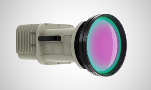
MKS sells Ophir Optics division to PCB maker TTM for $650M
October 22 2026


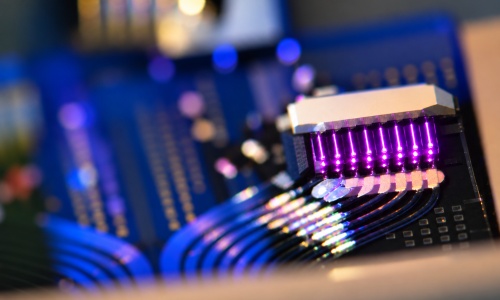
Photon Bridge lands €10M to scale lasers for data centers
October 21 2026