



Morgridge Institute reveals inner workings of immune cells
Autofluorescence lifetime imaging could improve diagnostics and offer new cell therapies.
02 September 2026
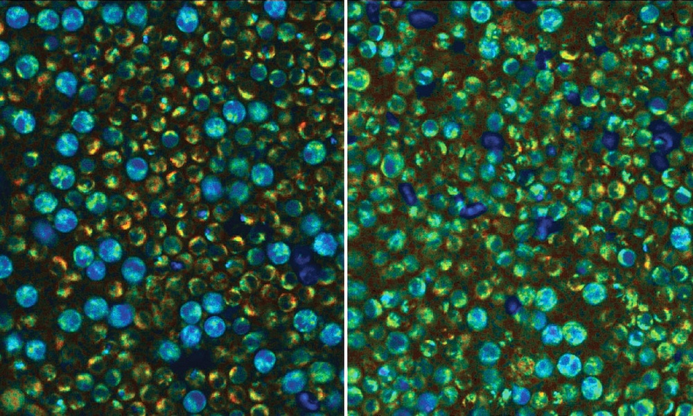
Metabolically quiescent (left) and activated (right) peripheral blood mononuclear cells captured with optical metabolic imaging Credit: Skala Lab/Jeremiah Riendeau.
A project at the US Morgridge Institute for Research has developed a new approach to the imaging of peripheral blood mononuclear cells (PBMCs), which are predominantly immune cells, as a route to understanding the inner workings of this type of biological component.
Published in Biophotonics Discovery, the findings could help improve diagnosis and treatment of immune system-related conditions like blood cancers, lupus, sepsis and cognitive decline.
Since PBMCs are also the starting material for CAR T-cell cancer therapies, which engineer a patient's own immune system to fight certain cancers, a better understanding of immune dynamics in these engineered cells could improve the outcomes of patients undergoing such treatments.
"PBMCs can be isolated clinically really easily, and they're already used in the clinical workflow," said Melissa Skala from the Morgridge Institute. "So the question is, what can we get from them that we aren’t already getting?"
The new study employed autofluorescence lifetime imaging, as a non-destructive alternative to existing techniques. Stimulated fluorescence from two particular dinucleotide molecules, termed (NAD(P)H) and FAD and both known to be metabolic cofactors in individual PBMCs, is sensitive to microenvironmental parameters such as pH, temperature and viscosity.
The Skala Lab fluorescence intensity and lifetime technique has been named optical metabolic imaging, or OMI, and was tested on samples of undifferentiated PBMCs isolated from donated blood of human volunteers. A 750-nanometer source excited the (NAD(P)H) nucleotides while 890 nanometers activated FAD.
"With OMI, a sensitive microscope shoots two long-wavelength photons, which don’t damage material as much as shorter-wavelength ones but have the same effect, at the sample," wrote the project. "This excites the inherent fluorescence of the products of cell metabolism and enables researchers to identify whether PBMCs are metabolically active or quiescent," ie. whether triggered to execute defensive action or in a resting state.
Making label-free metabolic information accessible to more labs
The results showed that that immune cells in the bloodstream are kept locked down in a quiescent state, noted the team, generally a desirable state when the system is not under attack. But when there is some infection or other problem, like cancer, this would cause an immune reaction and activate metabolic pathways.
"Having an understanding of the relative amounts of white blood cells is already used as a diagnostic biomarker," commented Morgridge's Jeremiah Riendeau. "Adding this metabolic piece can tell you additional facts about the activation state of the immune cells."
The Skala Lab believes its new type of analysis using OMI can help to make sense of heterogenous metabolic states between cell types, and how a subset of immune cells might be hyperactive while others remain quiescent. A less precise bulk measurement averaging across the whole sample would hide those differences, and obscure important data about whether cell therapy is successfully treating a cancer.
"Not every lab has access to a two-photon microscope, and so part of the research is making this label-free metabolic information accessible to more labs," said Riendeau. "Our lab and collaborators are in the process of commercializing our technology so we can make this measurement more available. The idea is that now that we understand how things look in a relevant sample, other labs can have this and do this, too."
Autofluorescence plus AI evaluates colorectal cancer in real time
September 02 2025
Tufts University observes Herpes virus reshaping neurons
August 26 2026
Diffraqtion aims high with $10M for 'quantum' camera platform
September 01 2026



SPIE O+P 2026: AI-based image analysis for bio-imaging
August 26 2026