


ICFO researchers refine Raman spectroscopy to better characterize melanin
Method reduces signal distortions enabling comparison of differently-pigmented samples.
24 June 2026
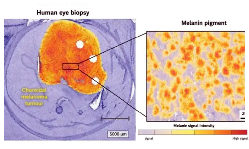
Human eye biopsy showing a choroidal melanoma (left) and a magnified view of melanin pigment within the tumour (right). Source: ICFO / Microchemical Journal.
Researchers at Barcelona-based photonics research center ICFO have developed a strategy that overcomes the challenges associated with Raman spectroscopy when characterizing the two main forms of melanin. The methodology, published this month in Microchemical Journal, reduces signal distortions and facilitates comparisons between differently pigmented samples and across varying experimental conditions. These results could guide future research on melanoma disease, say the researchers.
Melanin, the pigment that gives color to our skin, hair and eyes, is also believed to play a role in melanoma disease – the most dangerous form of skin cancer. To date, two main forms of melanin have been identified: eumelanin, which is photoprotective and antioxidant, and pheomelanin, which is phototoxic and prooxidant.
Correctly identifying and quantifying these could advance melanoma research, either by deepening the understanding of how the cancer works or by providing insights into effective treatment strategies. However, gold-standard methods for melanin characterization are destructive and require extensive sample preparation. Therefore, there is growing interest in developing non-destructive optical approaches for melanin analysis.
Non-destructive
Raman spectroscopy is one of the non-destructive optical approaches currently being explored for melanin analysis, as it provides molecular fingerprints of samples when illuminated by a laser. However, melanin analysis by Raman spectroscopy remains challenging due to the strong autofluorescence background and instrumental artefacts that can distort the measured spectra.
Now, ICFO researchers, José Javier Ruiz and Dr. Pablo Loza-Álvarez, Chief of the SLN Facility, together with Ismael Galván from the National Museum of Natural Sciences (CSIC), based in Madrid, Spain, have developed a strategy to overcome the challenges it presents.
The strategy – which was tested using hairs and feathers with different pigmentation – has shown that Raman imaging can reliably retrieve and characterize melanin information from intact biological samples.
There are two main difficulties that the proposal overcomes. First, it corrects a common distortion that complicates the interpretation of data, whose instrumental origin had not been clearly identified and corrected until now. “Several previous studies had observed a sinusoidal interference, which led to divergent results,” said José Javier Ruiz, first author of the article. “In our study, we describe how to characterize and correct it.”
Secondly, it addresses the broad and intense background signal caused by autofluorescence, which often masks the weaker Raman signals of interest. Rather than treating this autofluorescence solely as an unwanted interference, the proposal also takes advantage of it to provide complementary information about sample pigmentation.
“We anticipate that these advances may support future Raman and in general optically-based studies of different biological samples containing melanin, including research into melanocytic tumors”, said Dr. Pablo Loza-Álvarez, lead researcher of the study. “Improving the reliability and comparability of melanin spectral analysis, and making it more robust across different imaging platforms, could help to better understand how melanin composition and organization relate to different biological and pathological conditions.”

Exail poised for Thales takeover
July 13 2026
Magic Leap pivots to AR waveguide supply
July 13 2026