



Hyperspectral imaging maps scar tissue from heart attacks
George Washington University connects tissue damage to disrupted heartbeat.
05 August 2026
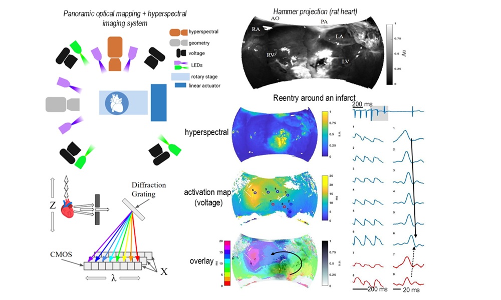
Hyperspectral mapping of tissue type and transmembrane potential. Credit: JBO.
Heart attacks leave behind injured muscle, which then turns to scar tissue. This scarring can in turn interfere with the electrical signals that coordinate each heartbeat, increasing the risk of arrhythmias.
Mapping electrical activity in the heart is straightforward, although seeing how those electrical signals relate to the scar tissue following a heart attack has proven more difficult.
Previous assessments of post-heart attack scarring have included the use of mechanical measurements, such as OCT imaging of hearts under elastographic testing; but a less invasive technique is more desirable, with in vivo assessment the ultimate goal.
A project at the George Washington University (GWU) has now demonstrated a platform combining hyperspectral imaging with optical mapping, to show where scar tissue is located and how electrical signals move around it. The findings were published in Journal of Biomedical Optics.
Optical mapping is a fluorescence imaging operation using voltage-sensitive probes to visualize fast changes in cardiomyocyte membrane potential; a powerful technique, although tissue type such as scar tissue is difficult to identify using optical mapping data alone.
GWU tackled this limitation by combining optical mapping with high-resolution panoramic hyperspectral imaging, using the spectral responses of cardiac tissues to illumination as a route to determining the tissue type at specific locations.
The researchers built an imaging platform employing six cameras and seven LED spotlights, to capture an ex vivo heart from multiple angles. Four cameras recorded electrical signals using a fluorescent dye that responds to changes in voltage, while one camera collected detailed spectral information from the tissue and one was used to reconstruct the heart's three-dimensional shape.
Illumination wavelengths were 365 nanometers for hyperspectral imaging and 520 nanometers for optical mapping, with specialized software combining all of the data into a single map of the heart surface.
Light signals from collagen
"No prior work has described a panoramic optical mapping system for imaging of electrical activity and co-registered panoramic hyperspectral mapping of tissue structure in living perfused hearts," wrote the project in its JBO paper.
In trials the imaging platform was applied to rat hearts four weeks after a heart attack had been induced. The hearts were removed, kept alive through a perfusion system, and scanned while researchers recorded both tissue properties and electrical activity.
The hyperspectral imaging component was able to identify scar tissue resulting from the heart attack thanks to the stronger light signal associated with collagen, which accumulates after cardiac injury. This allowed healthy heart muscle to be distinguished from damaged tissue, and the border zone between the two to be identified.
Using the combined approach let researchers see how electrical signals behaved near the scar. In several cases, abnormal beats originated close to the border between healthy and damaged tissue, while electrical waves often traveled rapidly around the scar but slowed down or became blocked when moving through scarred regions. These disruptions create conditions that support arrhythmias.
Since fibrosis, heart failure or aging can also alter heart tissue, the same approach could reveal new details of how the organ responds to a range of medical scenarios.
"This work demonstrates that panoramic hyperspectral mapping of tissue type and transmembrane potential is a powerful approach that enables functional mapping data to be analyzed within the context of local tissue type (healthy, infarct, and border) in living hearts," wrote the GWU team.
Johns Hopkins blood test detects earliest signs of heart attack
October 22 2024
OCE reveals tissue damage caused by heart attacks
January 29 2018

Battery welding pushes IPG sales momentum
August 04 2026


SPIE CEO Kent Rochford to retire in February 2027
August 04 2026


Multi-spot OCT tracks cornea deformations
July 29 2026