 |

15 Apr 2010
A MEMS scanning mirror designed for projection applications is at the heart of a handheld confocal microscope.
Researchers in the US have unveiled a handheld confocal microscope capable of capturing high-resolution images at video frame rates. The key to achieving this sought after combination is a MEMS scanning mirror developed for projection display applications. The team hopes that this breakthrough could pave the way towards a new generation of miniature confocal microscopes (Optics Express 18 3805).
"We achieve high-resolution images at 56 Hz, much faster than previous MEMS-based designs," Chris Arrasmith from Montana State University told optics.org. "Unlike other research that has targeted ultra-miniaturization, we wanted to demonstrate that the underlying MEMS scanner performance can support high fidelity imaging. Our results are the best in vivo skin images yet demonstrated using a MEMS scanner, and show that the MEMS scan technology is capable of diagnostic quality imaging. A future microscope designed around this scanner could be a much smaller pencil-like instrument."
Conventional confocal microscopes tend to be bench-top instruments lacking in portability. The goal of groups working in this field is to design smaller units that are both portable and handheld and can ultimately replace existing biopsy and histology methods for diagnosing suspicious skin lesions.
"Our entire unit, including control electronics, computer and light source, is mounted on a small cart that can easily be moved between exam rooms," commented Arrasmith. "The mobility of the instrument combined with the ability to be held and gather images in any orientation make it ideal for an in vivo diagnostic tool."
Confocal imaging of skin is a photon-starved problem so precise alignment and collection of all possible light is a must for high-quality images. The system designed by Arrasmith and colleagues David Dickensheets and Anita Mahadevan-Jansen uses an 830 nm laser diode and a dual-axis MEMS mirror that provides two-dimensional raster scanning of the laser beam to form the confocal image.
"Our system can image sub-cellular detail, such as cell membranes and nuclei," commented Arrasmith. "This is due to the use of high numerical aperture (NA>0.7) optics and a scan mirror that does not introduce any aberrations. We have also demonstrated imaging performance through the epidermis and into the dermis, as is evident from capillary blood flow visible in the videos obtained using the microscope. This is important for imaging diseases like melanoma, which begin at the dermal/epidermal boundary."
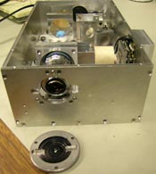
Having successfully completed the proof-of-principle stage of this work, the team is now looking to optimize the design and begin clinical trials. "The footprint of our first generation instrument is approximately 4 × 7 × 1.5 inch," said Arrasmith. "Part of this larger size was for experimenting with different objective lenses to achieve variable numerical aperture. In the generation 2 device, choosing a specific objective lens or designing custom optics will greatly reduce the overall size. It would be nice to see the next version measuring approximately 2 × 4 inch and 1 inch in height."
 |  |  |  |  |  |  |
| © 2025 SPIE Europe |
|