|

24 Jan 2007
The use of fluorescence measurement techniques is expanding rapidly, particularly in the life science and biotechnology sectors. Elaine Blackwood looks at what fluorescence lifetime has to offer compared with conventional measurement methods.
It is useful to define fluorescence from the outset. After absorbing a photon, a molecule can thermally relax to a metastable excited state, where it can emit a photon and return to the ground state. This emitted fluorescence photon has a lower energy than that of the absorbed photon that shifts the fluorescence emission towards the red spectral region, a so-called Stokes shift.
The fluorescence lifetime is the average time that the molecule remains in the excited state and can be thought of as a fingerprint of the electronic structure of the compound. In many instances, materials can be unambiguously identified by measuring the fluorescence lifetime.
It is also useful to state that fluorescence usually arises from decay from an excited singlet state with a typical lifetime in the nanosecond range. However, phosphorescence is associated with decay from an excited triplet state with a longer lifetime in the microsecond or millisecond range.
Steady-state fluorescence intensity measurements provide a typical increase of two orders of magnitude in sensitivity compared with conventional absorption spectroscopy, enabling samples of nanomolar to picomolar concentration to be analysed.
Single-photon counting techniques add a further order of magnitude improvement in sensitivity, the most sensitive instruments now measuring samples in the femtomolar concentration range.
However, fluorescence intensity measurements are only relative. In addition to being dependent on the excitation and emission wavelengths, the fluorescence signal depends on the intensity of the excitation source, the geometry of the measurement and the sample concentration. In contrast, the fluorescence lifetime is a self-referenced characteristic independent of excitation intensity and wavelength, as well as sample concentration.
The lifetime of a fluorophore is also affected by the microenvironment surrounding it. For example, the fluorescence lifetime can be quenched or enhanced by different photochemical processes, making it a useful environment sensor. This is the basis for using fluorophores as labelling probes in life science and biotechnology sectors, as well as in bio-assays.
A number of techniques have been developed to measure fluorescence lifetime. These include the phase-modulation method in the frequency domain and several techniques in the direct time domain, including time gating or the stroboscopic/boxcar method, streak cameras and time-correlated single-photon counting (TCSPC).
TCSPC is generally accepted as the method of choice as it gives direct measurement and observation of the decay kinetics in real time. It combines single-photon sensitivity and digital counting to give a high dynamic range with Poissonian noise statistics, enabling complex kinetics with long- and short-lived components to be analysed simultaneously from a single measurement.
In TCSPC, a pulsed light source repetitively excites a sample and the measurement builds a probability histogram relating the time between an excitation pulse and the observation of the first fluorescence photon.
By using a scattering reference sample, the instrument response function – which is broadened by contributions from the finite pulse width of the excitation source, the transit time spread of the detector and other electronic and optical effects – can be measured independently. Reconvolution software methods can eliminate the broadening due to instrumental effects and fluorescence lifetimes can be extracted to a higher precision. In practice, lifetimes down to one-fifth of the instrumental width can be determined accurately. TCSPC can typically measure lifetimes from a few picoseconds to microseconds.
Since TCSPC became commercially available almost 30 years ago, measurement times have improved from minutes or even hours to the present day, where high-quality data can be acquired and analysed in a little longer than the blink of an eye.
This major increase in data-harvesting speed has arisen due to the availability of short-pulse, high-repetition rate (up to 100 MHz) light sources (including mode-locked Ti:sapphire lasers, picosecond semiconductor diode lasers and modulated light-emitting diodes), short transit time single-photon counting detectors (photomultipliers, microchannel plate photomultipliers and single-photon avalanche diodes) and fast timing and data-acquisition electronics (often in the form of a PC plug-in card).
The actual measurement time depends on the precision required and, in turn, on the number of counts in the peak of the measured histogram and the number of individual time channels (typically from 256 to 4096). A reasonable estimate of an average lifetime can be obtained from only a few hundred counts in the peak measured in a matter of milliseconds, for example, from the emission of a single molecule detected in a confocal microscope. Higher-quality data with 103–104 counts in the peak is required to resolve complex kinetics and provide a precision in measurement lifetime to better than 0.1 ns, see figure 1.
Having established itself in the research laboratory, TCSPC is now a reliable, routine measurement, finding diverse applications limited only by the imagination of the user. Thanks to its sensitivity, TCSPC is currently being used for everything from single-molecule detection to lanthanide tracing; monitoring singlet oxygen in photodynamic therapy to studying carbon nanotubes and quantum dots; characterizing rare earth-doped glasses to bioscience areas such as protein folding, monitoring the reaction of enzymes and studying the dynamic structure of nucleic acids. New applications of fluorescence lifetime techniques have also emerged in biochemical assay development and high-throughput screening (HTS) for drug discovery.
Drug discovery
Fluorescence intensity (FI) detection techniques are now widely used in assay development and HTS where they are progressively replacing radiometric assay methods. Fluorescence detection avoids working with, and disposing of, radioactive materials. Other advantages include practically unlimited shelf life of labels, significantly increased stability of reagents and comparatively short measurement time.
Another major technical advantage is that fluorophores are highly sensitive to their environment, whereas radioactive isotopes provide little intrinsic information about their surroundings. Radioactive emission is characterized only by intensity, whereas fluorescence contains information in several parameters such as spectra, intensity, lifetime and polarization. This enables the design of multiparameter fluorescence-based assays, increasing the range of information obtained. These factors combine to make HTS by fluorescence techniques more cost-effective, rapid and amenable to miniaturization.
FI is currently the most popular readout parameter and is used in more than 30% of all screens. Measurements of FI require inexpensive instrumentation, but are often affected by interfering background signal from sample carriers, sample reagents, solvents and test compounds that could lead to false hits.
Time-gated fluorescence detection, often referred to as time-resolved fluorescence (TRF), is the second most popular fluorescence readout method. Time-gated detection requires pulsed sample excitation and uses the decay of long-lived (1–2 ms) lanthanide chelates and cryptates. Intrinsically an intensity measurement, its advantage over a steady-state FI measurement is that the short-lived (<10 ns) background signals and residuals of the lamp excitation pulse are removed by delaying the active detection gate with respect to the exciting light flash.
The goal of current HTS methods is to achieve higher-quality hits, essentially the ability to differentiate between true and false results, and reduce unnecessary cost and time for clinical trials. This puts increasing emphasis on the robustness of the assay technology and particularly the minimization of assay interferences. In this respect, fluorescence lifetime is ranked as the most robust homogeneous assay technology. However, its widespread acceptance has been delayed by the availability of suitable instrumentation.
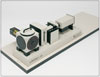
Bench-top fluorescence lifetime plate readers for homogeneous, liquid-phase-based assays are becoming available (see “NanoTaurus” image). Although specifically optimized for fluorescence-lifetime measurements, the system shown can measure other modalities like FI and polarization.
Direct measurement of the fluorescence lifetime of fluorophores within an assay offers the cost advantage of FI assays while matching, or indeed improving, the robustness and sensitivity of TRF assays. Such fluorophores have excited-state lifetimes in the nanosecond time regime, which are an intrinsic property independent of volume or concentration.
The ability of fluorescence lifetime to overcome interference is illustrated by measuring a series of samples of different concentrations of two dyes with individual lifetimes of 8.7 and 2.9 ns in solution with basic DMDM F12 cell culture medium (which also exhibits a lifetime of a few nanoseconds). These three components have significantly overlapping emission spectra that could not be separated by intensity measurements alone. TCSPC measurements analysed for the different lifetime components accurately reproduced their relative concentrations (see figure 2).
In conclusion, fluorescence lifetime measurements offer proven advantages in sensitivity and differentiation from interfering background emission and are set to become a leader in biotechnology assays and drug discovery.
 |  |  |  |  |  |  |
| © 2026 SPIE Europe |
|